Advanced Search
By: Sarah Plevin, BVMS, MRCVS, CVA, DABVP, ACVSMR
Overstrain injuries to the superficial digital flexor tendon (SDFT), which runs down the back of the canon bone, are among the most common musculoskeletal injuries 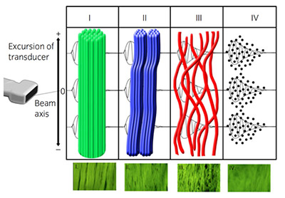 veterinarians diagnose in equine athletes. The many treatment options have three things in common: time out of training, expense, and no guarantee for success.
It makes sense, then, that injury prevention is always the goal; failing that, we need a method to guide rehabilitation.
Unfortunately, current imaging approaches limit how accurately veterinarians can monitor the tendon. So, although we can use conventional ultrasound to confirm the presence of a core lesion, we are often unable to fully evaluate tendon structure, follow healing, or provide effective rehabilitation guidelines post-injury.
A new ultrasound technology, however; called ultrasound tissue characterization could get us one step closer to achieving the goals of presenting injury and optimizing rehabilitation.
Ultrasound tissue characterization is relatively a new technique that might alleviate some of the problems we find with conventional ultrasound. It provides a 3D reconstruction of the tendon tissue and classifies it by color-coded echo types based on the integrity of the tendon ultrastructure (that which is only visible with an electron microscope). According to van Schie et al., this method allows practitioners to discriminate between healthy normal, adaptive/remodeling, and injured/healing tissue, often when conventional ultrasound findings are unremarkable.
The key to successive evaluations, allowing vets to look at tendon structure differences over time. Consecutive exams allow them to determine if a tendon is static, adaptive, healing, or degenerating and recommend changes in training intensity.
Researchers studying humans (Docking et al., 2015) and horses (Docking et al., 2012; Plevin et al., 2019) have reported ultrasound tissue characterization to be highly reproducible and user-independent. Their results suggest it's sensitive enough to detect the effect of changing loads (exercise) on tendons within days. This provides practitioners the opportunity to not only support rehab but also monitor tendons throughout training. Research is ongoing to determine whether the technology could help vets predict injuries.
Currently, physicians use it to monitor Achilles and patellar tendon health and guide exercise regimens post-injury in elite athletes. Vets use it in elite sport horses as part of routine tendon maintenance and to guide SDFT injury rehab.
What do the colors mean?
Green (Type 1 echoes) indicates normal, well-aligned, and organized tendon bundles.
Blue (Type 2) indicates wavy or swollen tendon bundles. This can represent remodeling tendon or inferior repair.
Red (Type 3) represents fibrillar tissue (the building block of tendon) from partial tendon rupture or initial healing.
Black (Type 4) are cells or fluid and represent core lesions where no normal tendon tissue exists.
How does it work?
A standard linear array ultrasound probe mounted on a motorized tracking device noninvasively and automatically moves down the SDFT over a 12-centimeter scanning distance. As it does, the device captures transverse images (horizontal planes) at regular distances and stores them in real-time for processing. The system records images every 0.2 millimeters to generate a tendon volume of 600 images. Image acquisition takes approximately 45 seconds.
Vets can subsequently use this volume to visualize the tendon in 3D to determine its structural composition and quantify tendon matrix integrity.
How can ultrasound tissue characterization help rehabilitation?
Scientists have shown that appropriate progressive loading helps stimulate tendon remodeling and healing - this is where this technique has the potential to be most useful.
By offering real-time information on tendon matrix integrity, ultrasound tissue characterization allows vets to take advantage of the limited window for appropriate tendon remodeling after injury. By mapping the tendon's ultrastructure and remodeling response to exercise at each step in the rehab regiment, ultrasound tissue characterization allows us to optimize the most vital tool in our rehab arsenal: exercise. Although research is ongoing, ultrasound tissue characterization gives vets a chance to adjust and tailor exercise regimens for individual tendons and horses, potentially allowing for a more successful return from injury.
Article provided courtesy of AAEP Media Partner, The Horse.